Cough Techniques for Tracheostomy
Cough Techniques for Individuals with Tracheostomy
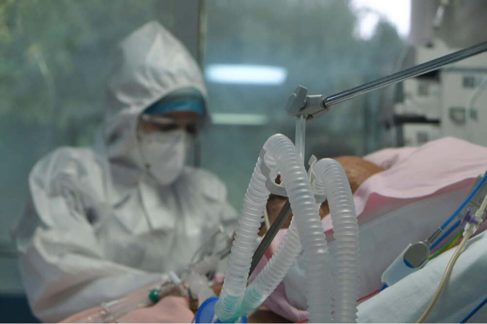
Patients with tracheostomy and mechanical ventilation have an impaired cough mechanism. Coughing is an important defense mechanism to remove irritants, pollutants, bacteria or any foreign objects (food, liquids, secretions) that have entered the airway. When material enters below the vocal folds and into the airway, this is termed aspiration. Coughing is a protective mechanism to help prevent material in the airway, and thus prevention of aspiration pneumonia.
A normal cough is initiated by the irritation of cough receptors which are found in the trachea, main carina, branching points of large airways, more distal smaller airways and are also present in the pharynx. Afferent signals are transmitted via the vagus nerve to the medulla, which then sends efferent impulses to the inspiratory, expiratory, and laryngeal muscles.
When anything obstructs the flow of air or irritates the respiratory tract, the vocal folds will open wider than usual, which will increase the amount of air being drawn into the lungs. There is then closure of the vocal folds, combined with contraction of muscles of chest wall, diaphragm, and abdominal wall which results in a rapid rise in intrathoracic pressure. During the expiratory phase, the vocal folds abruptly blow open resulting in high expiratory flow and the coughing sound. The cough is successful if enough pressure is generated to expel the material from the airway.
Since cough is an important defensive reflex, required to maintain the health of the lungs, individuals who do not cough effectively are at risk of atelectasis, recurrent pneumonia, and chronic airways disease from aspiration and retention of secretions.
Cough Reflex is Impaired with Tracheostomy
Individuals with tracheostomy will typically, at some point, have an impaired cough reflex. This may be due to the underlying medical condition. Patients with paralysis or neuromuscular disorders have a poorly developed and/or compromised cough reflex, and are rendered highly susceptible to lung infections and aspiration pneumonia. Those with obstructive lung diseases may not be able to generate the flow for an effective cough.
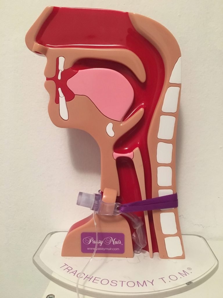
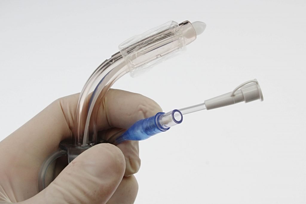
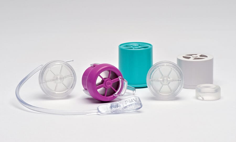
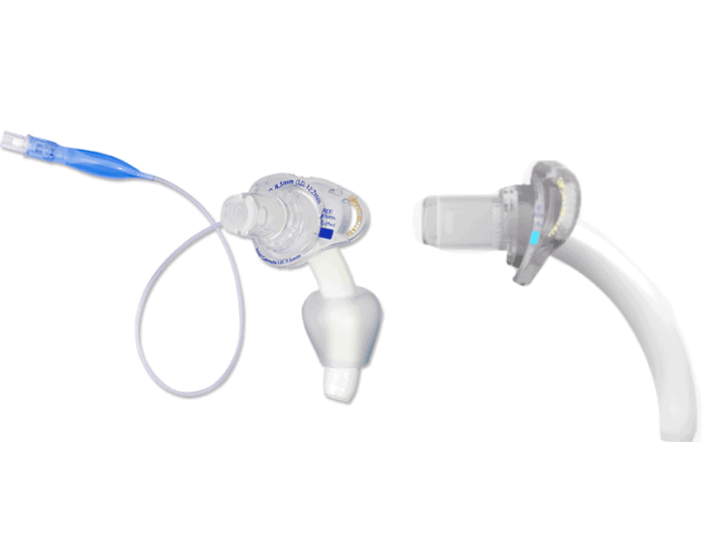
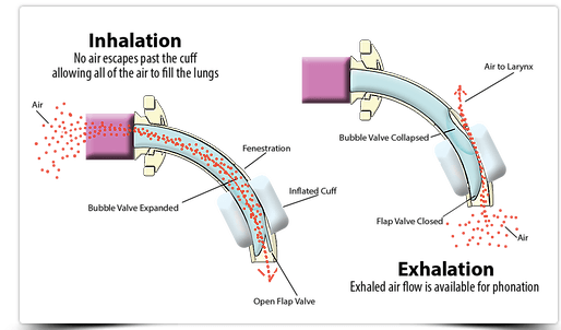
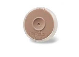
Individuals with tracheostomy, particularly with an inflated cuff, are unable to generate the pressure for an effective cough. With an inflated cuff, coughing results in expulsion of secretions through the tracheostomy tube, instead of the mouth. Secretions in the oropharynx and nasopharynx are unable to be cleared effectively. If the cuff is deflated, the patient will be able to generate more airflow through the upper airway. Following cuff deflation, occluding the tracheostomy tube such as with digital occlusion, a speaking valve or cap, enable the individual to use the upper airway and potentially produce an effective cough reflex.
The goal of airway clearance is to reduce airway obstruction, improve mucociliary clearance, improve ventilation and optimize gas exchange. For the patient with a tracheostomy, airway clearance is an important piece to decannulation.
To improve coughing in individuals with tracheostomy, some techniques include mechanical insufflation-exsufflation, manually assisted cough, and breath stacking or lung volume recruitment (LVR). Consult the doctor prior to providing these coughing techniques.
Mechanical Insufflation-Exsufflation
The mechanical insufflator-exsufflator (MIE) is a portable electric device that attempts to simulate a cough by applying a positive pressure to fill the lungs and then a negative pressure to provide a high expiratory flow rate to stimulate a cough. The device can be applied to the tracheostomy tube to assist a patient with an ineffective cough in clearing retained bronchopulmonary secretions.
Individual may need assistance with removing secretions above the cuff, particularly those with weak oropharyngeal musculature. In a study of high cervical spinal cord injury, a pharyngeal clearance manuever was utilized with a mechanical insufflator-exsufflator to improve secretion management. The Pharyngeal Clearance Maneuver (PCM) utilizes a modified application of the MIE device to mobilize “secretion burden” at the portion of the trachea above the tracheostomy cuff during cuff deflation. “Utilization of this strategy may reduce the risk of aspiration, infection, and respiratory compromise for patients with high cervical SCI in the acute rehabilitation setting” (Ehsanian, R et al, 2019). It can be most useful for individuals with large amounts of secretions, impaired coughing, and those whose cuffs are being deflated for the first time.
Mechanical insufflation-exufflation has been reported as more comfortable and effective than suctioning for individuals with tracheostomy. It is also more effective than manually assisted cough. In a randomized controlled trial, Pillastrini and colleagues verified an increase in peak cough flow. Other studies and a systematic review have also verified increased peak cough flows, and it increased above 270 L/min (level 1). Furthermore, MIE improves oxygenation in COPD and NMD patients (Arcuri, J et al 2016).
The use of MIE associated with non-invasive ventilation may prevent, delay or allow the removal of tracheotomy in patients with neuromuscular disease (level 2), decrease respiratory complications and hospitalizations (Arcuri, J et al 2016).
MIE is contra-indicated when patients present with increased risk of pneumothorax, which might happen in patients with some cancers, such as sarcoma, lung carcinoma, germ cell tumor or lymphoma (Arcuri, J et al 2016).
Manually Assisted Cough
Manually assisted cough is often performed as an abdominal thrust or lateral coastal compression.
In an abdominal thrust, the patient is best positioned either in a seated position or semi-lying with the head of the bed slightly elevated. The steps include the following:
- Place index fingers on the individual’s hip bones
- Slide the thumbs towards the belly button
- Place the heel of one hand, one inch above the belly button
- Place the other hand on top of the first hand with fingers interlocked with straight elbows, and fingers away from ribs or chest.
- Once the patient is in position, as him/her to take a deep breath or add as much air as possible to your lungs and hold the breath
- Cue to “cough strong.” As the patient is coughing apply one quick, forceful push inward and upward through the abdomen.
- Cue to expel the secretions out the mouth and remove with a tissue or suction.
This compensates for diaphragm weakness and drives the diaphragm upward.
Do NOT use abdominal thrusts with patients who have abdominal aneurysms, acute bleeding ulcers, pregnancy or recent abdominal surgery.
Lateral coastal compression can be used if abdominal thrusting is not possible.
- place the hands on the lower ribs with fingers pointing to the back.
- Have the patient take a deep breath and add as much air as possible into the lungs.
- Cue to “cough strong”, and squeeze the ribs up and in.
- Cue to expel the mucus out the mouth and remove with tissue or a suction.
Breath Stacking or Lung Volume Recruitment (LVR)
Breath stacking is a technique to increase lung inflation to clear mucous and improve cough effectiveness. The technique relies on glottic closure or, if necessary, a one-way valve to “hold in” sequential breaths. Since glottic closure is necessary, the tracheostomy tube must be occluded during exhalation, which can be achieved with digital occlusion, a speaking valve or cap. During mechanical ventilation, a speaking valve would be the only option to allow for positive pressure ventilation during valve use. Digitial occlusion, a speaking valve or cap may be used for individuals who are spontaneously breathing with tracheostomy.
Breath stacking can significantly improve peak expiratory flow and cough effectiveness by increasing lung volumes, increasing the elastic recoil pressure of the respiratory system, and optimizing the length of the expiratory muscles.
Technique
- Sit upright in a comfortable position
- Take a deep breath in and hold it
- Try to take another deep breath in on top of the previous breath
- Aim to take another 1-3 breaths in the same way (3-5 breaths in total)
- Breathe out or cough
- Repeat this process 3-5 times
The patient or a careprovider can also use a resuscitation bag to assist sequential spontaneous breaths, which are each followed by glottic closure. This technique may increase lung volumes and improve coughing by recruiting underinflated alveoli and collapsed lung segments, clearing secretions, and improving compliance and range Of motion of the lung and chest wall. Patients have anecdotally reported an ease in breathing, improved spontaneous cough efforts, and the sensation of a cleared airway for several hours after each LVR treatment session. It can help prevent atelectasis Results showed that LVR had a significant positive effect on forced vital capacity 15 minutes post treatment but did not have a facilitative effect at 30 minutes following treatment. LVR had a significant positive effect on peak cough flow (PCF) during unassisted coughing for up to 30 minutes (with a treatment effect size of .60). A moderate to large treatment effect in PCF was also observed 30 minutes after treatment across all post-swallowing clearance maneuvers including the supraglottic swallowing technique. The current rationale for doing LVR is to clear secretions, but if scheduled prior to meals, this treatment may provide enhanced airway protection and clearance over the length of a typical meal.
Inspiratory and Expiratory Muscle Strength Training
Inspiratory and expiratory muscle strength training may help to improve the cough and swallow function for patients with tracheostomy. Muscles for coughing may be weakened fro disuse atrophy when patients are debilated from acute or chronic disease. Atrophy can also occur when the cuff is inflated from disuse of the upper airway.
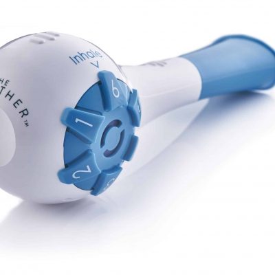
Inspiratory and Expiratory Muscle Strength devices such as the Breather, Acapella and EMST device have a place in helping to strengthen the pharyngeal and laryngeal muscles for coughing.
Free protocol for patients with tracheostomy and mechanical ventilation with purchase of The Breather.
Summary
Coughing is a protective mechanism which is impaired in individual with tracheostomy and mechanical ventilation due to the altered physiology with a tracheostomy tube. Coughing techniques for patients with tracheostomy can help to clear mucous and prevent atelectasis and pneumonia. These techniques are important for patients and careproviders to learn.
Resources
Arcuri JF, Abarshi E, Preston NJ, Brine J, Pires Di Lorenzo VA. Benefits of interventions for respiratory secretion management in adult palliative care patients-a systematic review. BMC Palliat Care. 2016;15:74. Published 2016 Aug 9. doi:10.1186/s12904-016-0147-y

