Anatomy and Physiology of the Aerodigestive Tract

All body systems are interrelated both anatomically and functionally.  This relationship is especially true for the aero-digestive system.  The aerodigestive system includes the upper respiratory tract, lower respiratory tract and digestive system.  An understanding of the normal respiratory and phonatory systems is necessary when working with tracheostomized and mechanically ventilated patients.  These systems are disrupted with the tracheostomy tube. 

The respiratory system consists of upper and lower respiratory tracts.  The upper respiratory tract consists of the nasal cavity, oral cavity, pharynx and larynx.  The lower respiratory tract includes the trachea, bronchi and the lungs. The digestive tract shares some of the same components as the upper respiratory tract.  

Upper Respiratory Tract

Nasal Cavity

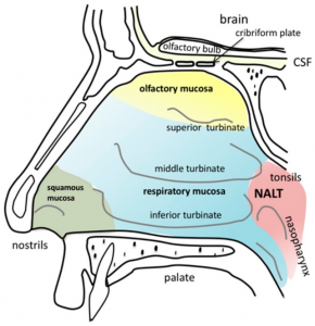
nasal cavity
Stella Gänger and Katharina Schindowski [CC BY 4.0 (https://creativecommons.org/licenses/by/4.0)]

The nasal cavity refers to the interior of the nose, or the structure which opens exteriorly at the nostrils. It is the entry point for inspired air and the first of a series of structures which form the respiratory system. The nasal cavity is involved in warming, humidifying and filtering air.  It also resonates sound and aides in our sense of smell and taste.  

The inside of the nose is lined with cilia or small hairs as well as mucous membrane.  Nasal hairs in the nares trap particles to prevent them from entering the respiratory tract.  The mucous membrane supports the body’s immune system.  It is sticky and helps to trap pathogens to protect against infection or allergies.  

Paranasal sinuses- The nasal cavity is surrounded by a ring of paranasal sinuses.  The sinuses develop as outgrowths of, and drain into, the nasal cavity. The mucosa of the sinuses connects to the nasal mucosa.

The lateral walls of the nose include three structures called the turbinates (inferior, middle, superior), which are finger like projections composed of bone and covered in soft tissue and mucosa.  The turbinates increase the mucosal surface of the nasal cavity and regulate nasal airflow.  Beneath each turbinate is a cleft or meatus.  

The nasal cavity is separated at midline by the nasal septum into the left and right side.  The nasal septum is composed of cartilage anteriorly, and bone posteriorly.  The bony septum is made up of the perpendicular plate of the ethmoid bone, vomer bone and maxilla bone.  The right and left nasal cavities become one at the choana, which is the opening of the nasopharynx. The conchae (turbinate bones) of the nasal mucosa expand the total surface area of the mucosa and create turbulence for air entering the respiratory passage. This causes air to swirl as it moves through the nasal cavity and increases contact between infiltrating air and the nasal mucosa, allowing particles in the air to be trapped before entering other parts of the respiratory system.

The nasopharynx contains a collection of lymphoid tissue called the adenoids.  The Eustacian tubes each open into the sides of the nasopharynx, connecting the middle ear with the upper respiratory tract.  


Oral Cavity

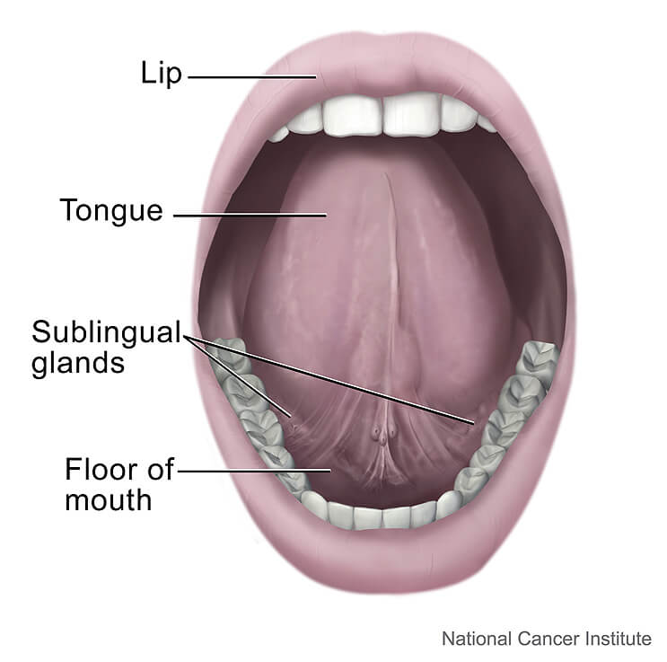
oral cavity

The oral cavity is separated from the nasal cavity by the hard palate (roof of the mouth).  The oral cavity incluldes the lips, front two thirds of the tongue, gums, teeth, buccal mucosa, floor of the mouth, retromolar trigone (area behind wisdom teeth), and hard palate.  The oral cavity is separated from the nasal cavity by the hard and soft palate.  The palate is the roof of the mouth and the floor of the nasal cavity.  The hard palate is the anterior two thirds and the soft palate is the posterior third.  

The lips seal the oral cavity which creates a pressure gradient for both speech and swallowing.  For speech, the lips are important for making bilabial sounds, /m, p, b/.  For swallowing, the lips form a seal so that food and liquids are not spilled anteriorly.  

The tongue is used for preparing food into a cohesive ball or bolus.  The tongue is also an important articulator, used to shape speech sounds.  

The oral cavity plays a significant role in respiration and it is also important for swallowing, digestion and speech. 

Pharynx

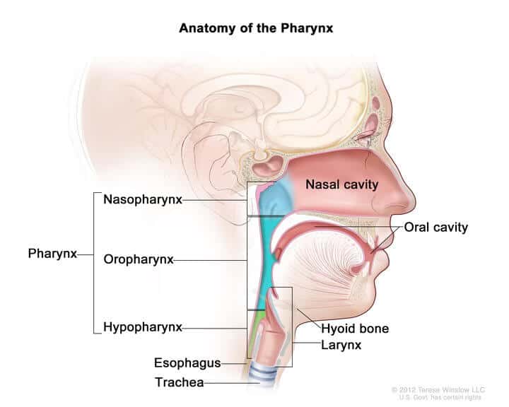
pharynx

The pharynx is situated immediately posterior to the nasal cavities and extends to the upper digestive tract.  The pharynx consists of the nasopharynx, oropharynx and hypopharynx. The nasopharynx extends from the base of the skull to the superior surface of the soft palate.  The oropharynx extends from the soft palate to the hyoid bone. The hypopharynx extends from the hyoid bone to the inferior aspect of the cricoid cartilage. 


The oropharynx includes the posterior third of the tongue (base of tongue), vallecula, palatine tonsils and tonsillar fossa, faucial arches, retromolar trigone, pharyngeal walls of the superior and lateral pharynx, soft palate, and uvula.  

The pharynx is a dual passage for respiration and swallowing.  It serves as a conduit for moving air to and from the lungs.  The pharynx also functions to transport boluses of food and liquid from the oral cavity and into the esophagus and stomach.  The pharynx must coordinate respiration and swallowing.  The palatal muscles, the extrinsic tongue muscles, and the hyoid muscles maintain the patency of the pharyngeal airway. 

A gag reflex may be induced by touching of the base of the tongue, soft palate, uvula, or posterior pharyngeal wall.  The gag reflex is considered a protective reflex to prevent foreign material from entering into the pharynx.  The presence of a gag reflex varies among the normal population with up to 37% having an absent gag reflex (Davies et al, 1995).  Therefore, lack of a gag reflex is not an accurate predictor of dysphagia (difficulty swallowing).  

Muscles of the Pharynx:

The pharyngeal muscles contract pushing the food into the esophagus.  There are two muscular layers of the pharynx: the outer circular layer and the inner longitudinal layer. The outer circular layer includes:

  • Inferior constrictor muscle
  • Middle constrictor muscle
  • Superior constrictor muscle

During swallowing these muscles constrict to propel the bolus downwards (an involuntary process).

The inner longitudinal layer includes:

  • Stylopharyngeus muscle
  • Salpingopharyngeus muscle
  • Palatopharyngeus muscle

During swallowing, these muscles act to shorten and widen the pharynx.  

The pharyngeal muscles are innervated by the pharyngeal branch of the vagus nerve (CN X) with the exception of the stylopharyngeus muscle, which is innervated by the glossopharyngeal nerve (CN IX).  


Vallecula

The vallecula is the space between the base of tongue and the epiglottis.  It is a common landmark during intubation.  

Soft Palate

The soft palate is a mobile portion made of up fiber and muscle that is suspended by the hard palate and ending at the uvula.  During swallowing, the soft palate elevates to contact the lateral and posterior walls of the pharynx, to prevent bolus regurgitation into the nasal cavity.  

Epiglottis

The epiglottis is a flap of tissue located at the base of the tongue.  It separates the respiratory and digestive systems for airway protection.  The epiglottis typically tilts downward during deglutition due to elevation of the larynx and approximation of the hyoid bone and thyroid cartilage (Ekberg, O et al, 1982).  It acts as a rudder to deflect food laterally, away from the airway to help protect the airway from aspiration.  In a case series, patients with an isolated epiglottectomy, were able to functionally swallow without aspiration (Leder, et al, 2010). 

Larynx

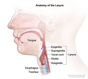
larynx

The larynx (voice box) is located between the pharynx and trachea. By adulthood, the larynx descends inferiorly.  The larynx functions in breath control,  voice production, and protection of the airway against aspiration.  The larynx houses the vocal folds.

The larynx is made up of three large unpaired cartilages:  epiglottis, thyroid, and cricoid.  It is also comprised of three smaller paired cartilages: arytenoids, corniculate, and cuneforms. 

Epiglottis

The epiglottis is one of nine cartilaginous structures that make up the larynx.  During breathing, it lies completely within the larynx. During swallowing, it serves as part of the anterior of the pharynx.  The depressions between the base of tongue and epiglottis are called valleculae.  

Thyroid cartilage:

The thyroid cartilage is the largest of the laryngeal cartilages.  It predominates anteriorly and forms the laryngeal prominance (Adam’s apple), which is more prominent in males.  The thyroid cartilage has superior and inferior horns.  Inferior horns make a synovial hinge joint with the cricoid cartilage. 

Cricoid cartilage:  

The cricoid cartilage is a complete ring of cartilage that surrounds the trachea.  The cricoid provides important attachments for muscles and ligaments.  It is located inferior to the thyroid cartilage, which it connects by the medial cricothyroid ligament.  The three paired cartilages (arytenoids, corniculate, and cuneforms) attach superiorly to the cricoid cartilage.  

Arytenoid cartilage

The paired arytenoid cartilages are found on the dorsal aspect of the larynx, attached superiorly to the cricoid cartilage. Both arytenoid cartilages give off a lateral extension (muscular process) and anterior extension (vocal process) which aid in supporting the vocal ligaments. Additionally, each arytenoid cartilage has an associated corniculate and cuneiform cartilage.

Corniculate cartilage

The corniculate cartilages are 2 small, conical cartilages that articulate with the apices of the arytenoid cartilages,  They play a role in opening and closing of the glottis, along with the arytenoids. 

Cuniform cartilage

The cuneiform cartilages are 2 small, club-shaped cartilages that lie anterior to the corniculate cartilages in the aryepiglottic folds.

Hyoid Bone

The hyoid bone is not technically part of the larynx.  The hyoid bone is a U-shaped bone situated anterior in the neck, between the chin and the thyroid cartilage.  It is the only bone that does not articulate with any other bone.  It can be palpated above the thyroid cartilage.  Pharyngeal muscles attach to the hyoid to elevate the larynx up and pull it forward. 

Nerve Supply to the Larynx

The larynx is innervated by the vagus nerve (X).  The vagus nerve is the longest nerve in the autonomic system in the human body.  
There are two branches: 

  • Superior laryngeal nerve
  • Recurrent laryngeal nerve
 Recurrent laryngeal nerve
 
The recurrent laryngeal nerve provides motor innervation to the intrinsic muscles of the larynx (except the cricothyroid). The paths of the left and right recurrent laryngeal nerves vary slightly with the left recurrent laryngeal nerve dividing from the main vagus nerve at the level of the aortic arch. The left recurrent laryngeal nerve then loops posteriorly around the left aortic arch to ascend through the superior mediastinum to enter the groove between the esophagus and trachea.  The right recurrent laryngeal nerve divides from the main vagus nerve at the level of the right subclavian artery to enter the superior mediastinum. The right recurrent laryngeal nerve then loops posteriorly around the right subclavian artery to ascend in the groove between the esophagus and trachea.
The recurrent laryngeal nerve branches into the inferior laryngeal branch.
 
  • Inferior laryngeal branch of the recurrent laryngeal nerve:  the inferior laryngeal branch of the recurrent laryngeal nerve innervates all intrinsic muscles of the larynx, with the exception of the cricothyroid muscle (innervated by the superior laryngeal nerve).
  • The recurrent laryngeal nerves also carry general visceral sensory fibers from the region inferior to the glottis.
  • The recurrent laryngeal nerve also sends branches to the inferior constrictor and cricopharyngeus muscles prior to entering the larynx
Risk to the recurrent laryngeal nerve is high in cases of thyroidectomy. Unilateral nerve damage presents with voice changes, including hoarseness. Bilateral nerve damage may result in aphonia (inability to voice) and breathing difficulties, with the potential need for a tracheostomy.
 
Superior laryngeal nerve
 

The superior laryngeal nerve arises from the inferior ganglion of the vagus.  At the level of greater horn of the hyoid, the superior laryngeal nerve divides into a smaller external laryngeal nerve and a larger internal laryngeal nerve.


Muscles of the Larynx

The larynx is composed of intrinsic and extrinsic muscles. 

Intrinsic Muscles: 

The intrinsic muscles of the larynx are responsible for sound production and the movements of the laryngeal cartilages and folds themselves. Their attachments fall between laryngeal cartilages. With the exception of the transverse arytenoid muscle, these muscles are paired bilaterally.  

Extrinsic Muscles

The extrinsic muscles of the larynx are found in bilateral pairs and assists the larynx in gross movements.  The extrinsic muscles can generally be divided into two groups: elevators of the larynx and depressors of the larynx.  The suprahyoid muscles and the stylopharyngeus (a muscle of the pharynx) elevate the larynx, while the infrahyoid muscles depress the larynx. 

The suprahyoid muscles include the diagastric (anterior and posterior belly), mylohyoid, stylohyoid and geniohyoid.  The infrahyoids include the thyrohyoid, omohyoid (inferior belly), sternohyoid, and sternothyroid. 

 The suprahyoid and infrahyoid muscle groups attach to the hyoid bone, which is bound to the larynx by strong ligaments, allowing the larynx to be moved as one unit.


Vocal Folds

Vocal folds are located within the larynx  at the top of the trachea. They are attached posteriorly to the arytenoid cartilages, and anteriorly to the thyroid cartilage. The vocal folds are open during inhalation, and close when holding one’s breath.  During phonation, the vocal folds vibrate, modulating the flow of air being expelled from the lungs. 

If intubation is required, the endotracheal tube is required to pass through the vocal folds.  This may result in injury to the vocal folds or structures around the vocal folds.  When intubated, the vocal folds are in an open position and are unable to perform one of the main duties of airway protection.  

The vocal folds are a major defense in preventing food and liquid from passing into the airway.  During swallowing the vocal folds close to protect the airway from material entering into the airway and lungs.  When food passes below the vocal folds, this is called aspiration.  Aspiration can lead to inflammation and possible pneumonia.  Patients with tracheostomy are at high risk for aspiration.  See the swallowing management section for more information.  

The supraglottis, glottis and subglottis are the three anatomic subsites of the larynx. 

Subsites of the larynx

Glottis

 The glottis is the space between the vocal folds.  Intubation may cause injury to the glottis due to pressure from the arytenoid cartilages.  

Subglottis

The subglottis or subglottic region is an anatomical area of the larynx that is located inferior to the glottis.  

The subglottic region is also an area of injury due to intubation.  Pressure or movement of the endotracheal tube can cause necrosis.  Tracheal stenosis may also occur in the subglottic region from intubation or due to complications of a tracheostomy cuff.  See cuff management for more information. 

Supraglottis

The supraglottis or supraglottic region is an anatomical area of the larynx that is located superior to the glottis.  The superior extent of the supraglottis is the tip of the epiglottis, and the supraglottis extends inferiorly to the laryngeal ventricle (which separates the false from the true vocal cords).  The supraglottis includes the epiglottis, false vocal cords, aryeptiglottic folds, and arytenoid cartilage.

Esophagus

The esophagus is a muscular organ that allows for the passage of food into the stomach.  The esophagus begins at the upper esophageal sphincter (UES).  The UES is a muscular band that relaxes to allow food to enter the esophagus when the larynx elevates & pulls forward.  

Lower Respiratory Tract

The lower respiratory tract is the part of the respiratory system from the trachea to the lungs. There are 16 to 20 C-shaped cartilage rings that serve to support the trachea to keep it patent during respiration. A tracheostomy incision is usually cut between the first and second or second and third tracheal ring.

The posterior  trachea and the anterior esophagus share a common muscular wall that lacks  cartilaginous structure.  This smooth muscle allows the esophagus to bulge into the tracheal wall during a swallow, permitting the food bolus to pass to the stomach.  

The trachea bifurcates into the right and left mainstem bronchus. The angle of the right mainstem bronchus is less acute than the angle of the left, therefore the right mainstem bronchus is more susceptible to aspirated material.  However, this is dependent on patient positioning as well.  

The bronchi divides further into secondary and tertiary bronchi, which continue to branch to smaller airways called the bronchioles. The bronchioles end in air sacs called the alveoli.  The alveoli are tiny balloon shaped structures that are the smallest passageway in the respiratory system, that are arranged in clusters in the lungs.  There are about 600 million alveoli in healthy lungs.  They allow for gas exchange to occur.  Alveoli are lined by a fluid layer known as a surfactant, which maintains the shape and surface tension of the air sac.

There are many medical conditions that can effect the alveoli and cause the alveoli to become inflated and/or scarred.  These conditions include emphysema, pneumonia, tuberculosis, acute respiratory distress syndrome (ARDS), and pulmonary edema.  

The lower respiratory tract is not involved in digestion.  If food or liquid reaches the lower respiratory tract, the term used to describe this is aspiration.  Aspiration is when any foreign material (food, liquid, secretions, reflux) enters the airway below the level of the vocal folds.  Aspiration pneumonia is a complication that can occur from aspiration.  Not every patient who aspirates will develop pneumonia.  Individuals who have poor general health and poor oral care are at highest risk of developing pneumonia due to aspiration.  

Lungs

The lungs are a vital part of the respiratory system.  The lungs are a pair of spongy, air-filled organs located on either side of the thorax (chest).  They divide into smaller segments called lobes.  The right lung consists of three lungs and the left lung is slightly smaller containing two lobes.

The lungs are covered by two thin tissue layers called pleura, which protect and cushion the lungs .  The visceral pleura (inner layer) covers the lung surface.  The outer layer (parietal pleura) lines the inside of the chest wall. The very thin space between the layers is called the pleural cavity.  A liquid, called pleural fluid, lubricates the pleural cavity so that the two layers of pleural tissue can slide against each as the lungs inflate and deflate during breathing.  There is no anatomical connection between the right and left pleural cavities. A pathological condition of the pleural fluid is called pleural effusion.  

Intrapleural pressure

Intrapleural pressure or intrathoracic pressure is the pressure within the pleural cavity.  Normally, the pressure within the pleural cavity is slightly less than the atmospheric pressure, in what is known as negative pressure.  This negative pressure acts like a suction to keep the lungs inflated.  The negative pressure is due to three main factors:  

  1. The surface tension of the alveolar fluid- The surface tension of the alveolar fluid tends to pull each of the alveoli inward and therefore pulls the entire lung inward. Surfactant reduces this force. 
  2. The elasticity of the lungs- The abundant elastic tissue in the lungs tends to recoil and pull the lung inward. As the lung moves away from the thoracic wall, the cavity becomes slightly larger. The negative pressure this creates acts like a suction to keep the lungs inflated. 
  3. The elasticity of the thoracic wall- The elastic thoracic wall tends to pull away from the lung, further enlarging the pleural cavity and creating this negative pressure. The surface tension of pleural fluid resists the actual separation of the lung and thoracic wall.
Intrapleural pressure changes during breathing.  As the thoracic wall moves outward during inspiration, the volume of the pleural cavity increases slightly, decreasing intrapleural pressure. As the thoracic wall recoils during expiration, the volume of the pleural cavity decreases, returning the pressure to minus 4, or 756 millimeters of mercury.  
 
A pneumothorax, or collapsed lung, occurs when air leaks into the pleural space.  There is no anatomical connection between the right and left lungs, each surrounded by its own pleural cavity and pleural membranes. Therefore, changes in the intrapleural pressure of one lung do not affect the other lung.

Muscles of Respiration

There are inspiratory and expiratory muscles of respiration to help with breathing.  The inspiratory muscles include the diaphragm, intercostals, pectoralis major, pectoralis minor, strap muscles of the neck, sternocleidomastoid, and scalene muscles. Muscles of expiration include the abdominal and the internal and external intercostals. 

Events during Quiet Inspiration

The primary muscle of inspiration is the diaphragm.  The diaphragm is a dome shaped muscle in the resting position.  During inspiration, the diaphragm and external intercostal muscles contract.  The diaphragm moves inferiorly, increasing the volume of the thoracic cavity. This causes the intrapleural pressure to become more negative, which increases the transpulmonary pressure, causing the lungs to expand. The expansion of the lungs lowers the intrapulmonary pressure below atmospheric pressure. Air, following its pressure gradient, now flows into the lungs.


Events during Quiet Expiration

During expiration the muscle returns to the relaxed, dome shaped position.  During expiration, the diaphragm and external intercostal muscles relax, decreasing the volume of the thoracic cavity. The intrapleural pressure becomes less negative, the transpulmonary pressure decreases, and the lungs passively recoil. This increases the intrapulmonary pressure so that it rises above atmospheric pressure.  Air, following its pressure gradient, moves out of the lungs. 

Accessory Muscles

The pectoralis major and minor, strap muscles of the neck, sternocleidomastoid and scalene muscles are all accessory muscles of inspiration used during forced breathing such as physical exertion. 

References:

Davies AE, Kidd D, Stone SP, MacMahon J (1995). Pharyngeal sensation and gag reflex in healthy subjects. Lancet 345:487–488. 
Google Scholar | Crossref | Medline | ISI

Ekberg, O, Sigurjónsson SV. (1982). Movement of the epiglottis during deglutition. A cineradiographic study. Gastrointestinal Radiology. 7(2):101-7. 

.

join trach courses

Subscribe now