Ten Communication Options for Individual with Tracheostomy and/or Mechanical Ventilation

Individuals with tracheostomy and mechanical ventilation with an inflated cuff are unable to produce voicing, and many do not speak for an extended period of time.  Normal voicing occurs because airflow generated from the lungs passes through the vocal folds, resulting in vibration of the folds, which produces voicing.  When a patient has a cuffed tracheostomy, the airflow is redirected out the tracheostomy tube.  If no airflow passes through the vocal folds, the individual will be unable to voice.  These patients have difficulty expressing basic wants and needs and difficulty participating in life and death healthcare decisions.  One study found that 33% of conscious, ventilated intensive care unit (ICU) patients were unable to communicate with caregivers (Zubow & Hurtig, 2013).  Happ et al, 2015 reported that 53.9% of their mechanically ventilated ICU patients met their criteria for needing assistance with communication. Patients with Covid-19 may have even more difficulty with communication due to masks being worn which distort the staffs communication attempts.  

Numerous methods can be used to communicate including gestures, head nods, writing, use of communication boards, augmentative communication.  These methods may be tailored to meet individualized patients’ needs.  Leak speech, speaking valves and talking tracheostomy tubes are methods for the individual with tracheostomy to produce voicing.  

A communication assessment should begin prior to the procedure when nonemergent tracheostomy is planned (Mitchell et al, 2013).  Speech-language pathologists are typically trained in finding the most advantageous communication method for patients with tracheostomy. 


Consequences of being unable to communicate

Individuals with tracheostomy and/or mechanical ventilation often have difficulty with one of the most basic human functions, communication.  The inability to effectively communicate with medical staff and other care providers during an acute illness makes it difficult for patients to understand their illness, participate in medical decision making, and be active in their treatment.  Communication is particularly critical in health care environments where miscommunication may lead to misdiagnosis or delayed medical treatment.  Patients often report frustration, fear, powerlessness and anxiety due to loss of voicing (Tolotti, A et al 2018).  

Patients with communication disorders have a higher rate of medical complications and errors (Barlette et al., 2008) and are at increased risk of preventable adverse events (JACHO, 2010). Adverse events lead to poorer patient outcomes, unnecessary patient suffering and dissatisfaction, longer hospital stays, and extra healthcare spending each year (JACHO, 2011).  Hospitals in the US, seeking Joint Commission accreditation, must adhere to standards intended to ensure that health care providers communicate appropriately and effectively with patients.

Joint Commission has provided a Roadmap for Hospitals for Advancing Effective Communication, Cultural Competence, and Patient and Family Centered Care. 

Not only is communication mandated and a patient right, but it is also an ethically responsibility.  Re-establishing communication for individuals with a cuffed tracheostomy tube has a significant impact on quality of life ( Khalaila, R et al, 2011; Stein-Parbury J, & McKinley S., 2000). 

There are also developmental risks for young children with a tracheostomy.  Young children with tracheostomies do not get to explore making sounds. They also may have limited social interactions that are critical to the development of language skills.  Caregivers tend to talk less to children who cannot communicate.  Children are at risk for delays if therapeutic intervention is not initiated. 

Emergency Call Systems

When an individual is in a medical setting, an emergency call system is important for patient safety and a means for patients to communicate with their care providers to have their needs met.  There are a variety of call bell systems.  Technology has evolved to two way audio systems where nurses can communicate with a patient about their needs in their room before responding to the call.  Systems can also integrate with wander management, security systems, and fire alarms.  Faster response times to calls have resulted in reduced fall rates and reduced injurious fall rates (Tseng et al, 2012).  

Access methods depend on the abilities of the user, and may include the use of direct selection of symbols on the screen or keyboard with a body part, pointer, adapted mouse or joystick, or indirect selection using switches and scanning.

Augmentative and Alternative Communication Options

No Technology Options

No technology communication options for patients with tracheostomy and mechanical ventilation include gestures and sign language.  Gestures include responding to a communication partner with yes and no responses.  Facial grimacing may be a sign of pain.  Pointing is a technique to show a partner an item that is needed.  A limitation of gestures is that it limits the amount of information that can be provided.  

Establishing a clear yes and no is important for understanding a patient’s wants and needs.  This can be acheived by the patient nodding yes or no, blinking once for yes/twice for no, thumbs up or down, or eyes up and down.  

Sign language has a multitude of signs and is the method of choice in an institutional setting for individuals already knowledgeable of this system, such as those who are deaf.  It is a visually interactive language that uses a combination of hand motions, body gestures, and facial expressions.  Sign language is only functional if the communication partner understands the signs being communicated.  There are sign language interpreter systems that facilities may rent or purchase to make available to help with communication needs of those whose primary language is sign. 

Low Technology Communication Options

Low-tech communication aids are defined as those that do not need batteries, electricity or electronics. These are often very simple communication boards or books, from which the user selects letters, words, phrases, pictures, and/or symbols to communicate a message.  These solutions are typically for short term use.  

If the user has the fine motor coordination and is capable, writing may be a low cost solution for a patient with a tracheostomy or mechanical ventilation to communicate.  This can be accomplished with a writing utensil and paper or marker and dry erase board.  However, writing is limiting because of the increased amount of time to get the message across, particularly for individuals in a medical setting.  The patient may also be too weak to write in the acute phase such as in ICU settings. 

Simple communication boards provide pictures or symbols to communicate.  Depending on physical abilities and limitations, users may indicate the appropriate message with a body part, light pointer, eye-gaze direction, or a head/mouth stick.  Alternatively, the user might indicate yes or no as a listener scans through possible options.  These types of communication boards limit the number of messages that the individual can communicate.  Alphabet boards may supplement communication, with the user pointing to different letters to create words.  

The E-tran is another low technology communication option for individuals with limited physical abilities, in which eye gaze is used to communicate.  The message is conveyed by the message sender selecting the desired component of the message using eye movement, while the communication partner deciphers the message by standing in front of them, tracking their eye gaze.

Communication boards have been shown to increase efficiency and speed of communication and decrease frustration of intubated patients (Hoorn et al, 2016). 

The Megabee is an easy to use communication device, that may benefit people with tracheostomy and mechanical ventilation who are unable to speak and unable to write legibly.

The tablet has been designed to require minimal learning for both communication partners.  Letter selection is achieved by looking at the letter and blinking, then looking at the block which is the same color as the selected letter and blinking again. The chosen letter will light up to indicate the appropriate letter and will be seen on the tablet.  The Megabee may be purchased through our Tracheostomy store, here.  

High Technology Communication Options

Communication Tracheostomy
©2019 Tobii Dynavox. All rights reserved

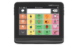
High-tech systems may be useful for patients with tracheostomy and mechanical ventilation, particularly if the patient is suspected to be long term ventilated and is unable to meet communication needs due to severe speech or language disorders.  Patients with degenerative disease, such as ALS, are likely appropriate candidates for high-technology communication.  There are dedicated devices developed solely for Augmentative and Alternative Communication (AAC) or non-dedicated devices such as computers that run additional software to allow the device to function as an AAC device. High-tech devices vary in size and weight, as well as how the user can access messages, including the use of direct selection of a screen or keyboard with a body part, pointer, or eye gaze, adapted mice or joysticks, or indirect selection using switches and scanning.

I 12 SCF English

High tech options may be static or dynamic in form.  Static communication devices have symbols in fixed positions with all the symbols constantly displayed on the device. On dynamic AAC devices, multiple pages of symbols are possible, and thus only a portion of the symbols available are visible at any one time, with the communicator navigating the various pages.

Oral Communication Options for Tracheostomy and Mechanical Ventilation

Oral communication is the preferred method of communication as it approximates normal communication.  Some patients are candidates for oral communication but also need another method of communication.  Options for oral communication include: 

Mouthing Words

There is no airflow or limited airflow though the vocal folds and the upper airway if an individual has a tracheostomy tube with an inflated cuff.  Natural speech requires airflow through the vocal folds to vibrate and produce voicing.  Therefore individuals are aphonic (no voicing) with an inflated cuff.  Many individuals attempt to mouth words to communicate basic wants and needs.  

To improve intelligibility and comprehension during mouthing of words:

  • Cue the individual to emphasize articulatory movements:  “Open your mouth large when speaking to enunciate each word.”  
  • Reduce the rate of speech
  • Use short phrases
  • Use simple words
  • Even if the resident is not on an oral diet, placing dentures can help improve the visibility of speech sounds for better intelligibility.  
  • Repeating each word the resident says is a technique for more accurate interpretation of the message.  
  • Encourage the individual to seek acknowledgement that the message has been understood. 

Although mouthing words is frequently employed with patients with tracheostomy and mechanical ventilation, there are many limitations.  There are many speech sounds that are not visible to lip read and not all communication partners are able to lip read.  The resident’s needs are often not accurately interpreted, which may lead to misdiagnosis or delayed treatment.  An example is a patient requesting for pants, and the nurse misinterprets as “pain”, and thus provides pain medication.  This is why it is important to repeat the patient and make sure the patient affirms that the message is received correctly.

Another negative of mouthing words is that the patient is unable to call for help in an emergency.


Electrolarynges for individuals with tracheostomy

Electrolarynges are typically used for individuals with laryngectomy.  However, they are also an option for individuals with tracheostomy and mechanical ventilation.  An electrolarynx is a battery operated device that is either placed on the neck or intra-orally.  The electrolarynx provides a tone which can be shaped by the articulators (tongue, lips, palate, teeth) into speech sounds.  The sound is often described as robotic and monotonous.  Results from a systematic review showed that 79-100% of individuals with tracheostomy were able to communicate effectively with an electrolarynx and was preferred over mouthing words, sign language and writing (Hoorn et al, 2016). 

Restoring Phonation in Patients with Tracheostomy and Mechanical Ventilation

Phonation is possible for patients breathing spontaneously with a tracheostomy tube as well as for those with tracheostomy on mechanical ventilation.  Medical complications of the pharyngeal, laryngeal, and tracheal structures, including glottic or subglottic edema, ulceration of the vocal fold, vocal fold paresis/paralysis, tracheal stenosis, and tracheomalacia, can affect the ability to vocalize.  

Phonation can be achieved in a few ways: Fenestrated tracheostomy tubes, leak speech, digital occlusion, speaking valves, talking tracheostomy tubes and the Blom Tracheostomy System.  

Fenestrated Tracheostomy Tubes

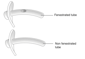
Fenestrated tracheostomy tubes have holes in the tube to allow for airflow through the tube when a fenestrated inner cannula is in place to enable voicing.  Individuals may voice when the cuff is inflated, partially deflated, or completely deflated.  

Fenestrated versus Non-fenestrated Tracheostomy Tubes

A drawback of a fenestrated tracheostomy tube is that the positioning of the fenestrations often rub against the trachea wall and can result in granulation tissue formation.  There is also a risk of subcutaneous emphysema when using a fenestrated tracheostomy during positive pressure ventilation (ICS, 2015).  A non-fenestrated inner cannula should be used during suctioning so that the catheter does not inadvertently pass through the fenestrations, resulting in trauma to the tracheal wall. 

Leak Speech

Again, an inflated cuff on the tracheostomy tube prevents airflow through the vocal folds and the upper airway.  Since the vocal folds require airflow to vibrate for voicing, individuals with an inflated cuff of the tracheostomy tube are unable to render speech.  

If the cuff is either partially or completely deflated, some airflow is likely to “leak” around the tracheostomy tube and through the vocal folds and upper airway.  Hence the term, leak speech.  There must be adequate space between the outer tracheostomy tube and the trachea for adequate exhaled gas to pass.  If the patient is in volume control, airway patency may be assessed by checking the exhaled tidal volume and the peak inspiratory pressure (PIP).  If the exhaled tidal volume and PIP significantly reduce following cuff deflation, then it is likely that the upper airway is patent.  If there is no voicing and PIP and exhaled tidal volume do not reduce significantly, even after full cuff deflation, check the tracheostomy tube size and determine if a downsized tube is appropriate for the patient to allow more airflow around the tracheostomy tube and through the upper airway.  Endoscopic assessment of the upper airway may also be indicated if airflow is not attained after deflating an appropriately sized tracheostomy tube.  

When a patient is on mechanical ventilation, leak speech can occur on both inspiration and expiration.  Since speech may be clearer when the ventilator delivers a breath on inspiration, individuals are frequently taught to speak when the ventilator delivers a breath.  However, this is the opposite of normal breathing and speech patterns and may result in dysphonia.  Another limitation of leak speech is that speech is often breathy and weak since airflow takes the path of least resistance, often back through the tracheostomy tube instead of through the vocal folds and upper airway. 

Education is important including coaching the patient on the expectation of airflow through the upper airway and how to coordinate speaking and breathing during cuff deflation.  Often during the initial cuff deflation, the patient may allow a large amount of air to escape through the upper airway, which can lead to shortness of breath, hypercapnia, or dryness.  

Ventilator changes with leak speech:

Ventilator settings may need to be adjusted by the respiratory therapist or physician to make sure the individual is ventilating adequately.  Ventilator changes may also be made to improve speech intelligibility and length of utterance.  Ventilator changes include increasing the set Positive End Expiratory Pressure (PEEP) and lengthening the inspiratory time.  If the PEEP is set to zero, most of the exhaled gas escapes out the tracheostomy tube, with little airflow through the upper airway for speech on exhalation.  Providing set PEEP when the cuff is deflated can increase the amount of utterances per breath.  Lengthening the set inspiratory time (iTime) can also provide longer speech as well as improve patient comfort. The set rate may also be a setting that can be increased during leak speech to improve speech output.  For example, if the ventilator is set at 8 breaths with an inspiratory time of 1 second, this will result in approximately 8 seconds of speaking time per minute.  If the set rate is 14 breaths per minute, with an inspiratory time of 2 seconds, this will allow for 28 seconds of speaking time per minute.  

Although changing the ventilator settings may improve speech output, it is important to make sure the patient is achieving adequate ventilation.  The higher respiratory rates require smaller tidal volumes to achieve PaCO2.  Prior to re-inflating the cuff, additional PEEP should be turned off to prevent hyperinflation.  All vent settings should return to settings prior to cuff deflation.  

 Low minute volume alarms are expected to alarm with full cuff deflation due to the escape of gas through the upper airway during cuff deflation.  Do not turn alarms off if the individual is not directly monitored by staff.  Since exhaled volumes are not fully returned to the ventilator with cuff deflation (some air passes through the upper airway), some alarms can be adjusted or silenced to stop unnecessary alarming.  The high and low pressure alarms should remain intact and adjusted appropriately to detect and alert caregivers to disconnects, patient fatigue, or changes in peak airway pressures.  An external low pressure alarm can be put into place if the ventilator does not have a low pressure alarm setting.       

Digital Occlusion

Tracheal occlusion using a finger is a technique to produce voicing for patients with tracheostomy who are spontaneously breathing only.  The cuff must be completely deflated or a cuffless tracheostomy tube must be used.  When the cuff is inflated, air flows in and out of the tracheostomy tube and air is prevented from escaping through the upper airway by the cuff.  If the cuff is inflated and the tube is occluded, air will not be able to escape out of the tracheostomy tube since it is occluded or through the upper airway due to the cuff, and therefore the individual would be unable to breathe.  

The procedure for digital/finger occlusion is as follows:

  1. Educate the individual with a tracheostomy about airflow and the steps for finger occlusion.
  2. Suction the patient as needed. 
  3. Slowly deflate the cuff of the tracheostomy tube and suction again if necessary. This step would be skipped for individuals with cuffless tracheostomy tubes. 
  4. Once the cuff (if present) is completed deflated, finger occlusion is performed by placing a gloved finger over the proximal opening of the tracheostomy tube, which redirects the airflow around the tracheostomy tube and through the upper airway.  Cue the patient to take a deep breath and occlude the tracheostomy tube on exhalation only.  Speech would be coordinated by placing the finger over the tube during speech on the exhalation cycle. 
 
The above procedure is also the assessment for airway patency to determine if an individual can breathe around the tracheostomy tube and through the upper airway.  If there is no voicing and limited airflow though the upper airway, check the tracheostomy tube size and determine if a downsized tube is appropriate to allow more airflow around the tracheostomy tube and through the upper airway.  It is critical to check airway patency  prior to placing a speaking valve or cap.  This method of checking for airway patency can be used for individuals who are spontaneously breathing with a tracheostomy tube.  An endoscopic assessment of the airway may also be warranted if there is any concern regarding airway patency.  
 
Limitations of finger occlusion are that it requires the dexterity to occlude the tracheostomy tube.  Infection control is also an issue.  If the finger or gloved finger has touched other objects in the room, this is a potential source for infection.

Speaking Valves

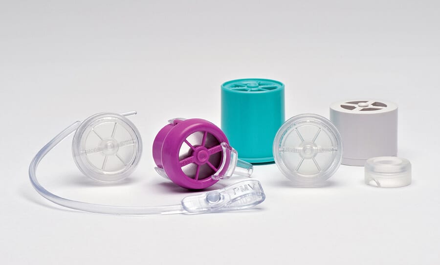
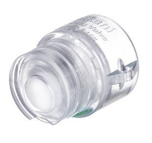
Speaking valves are one way valves that are designed to allow patients with tracheostomy the ability to produce natural voice.  Some speaking valves (Shiley, Shikani, Tracoe) can only be used for patients who are spontaneously breathing.  The only speaking valves approved for patients on mechanical ventilation are manufactured by Passy-Muir and Montgomery.   

All speaking valves are considered open-position valves except the Passy-Muir Speaking Valve, which is designed as a bias-closed position valve.  The bias-closed position means that the valve remains in a closed position at all times, opening only when the patient inhales.  At the end of inspiration, the Passy-Muir Valve returns to the closed position, creating a column of air inside the tracheostomy tube so that secretions do not build inside the tube.  All expired air passes around the tracheostomy tube (and deflated cuff if present), through the vocal folds and upper airway for speech.  The Passy-Muir Valve thus provides benefits other than speech by restoring a closed respiratory system including:  restores positive airway pressure, improved swallowing and may reduce aspiration, reduced secretions, facilitates weaning and decannulation.

An open position speaking valve has a diaphragm in an open position, meaning that expiratory flow is needed to close the diaphragm.  As a consequence the first part of expiratory air has to travel through the tracheostomy tube and close the diaphragm of the speaking valve, leaving the rest of expiratory air to flow through the upper airway. This can result in patients coughing secretions into the tracheostomy tube and partially blocking the open-position speaking valve.  Since there is a leak with open position valves, the system is not closed and positive pressure is unable to sustain.  To find out more about the differences of speaking valves and more in-depth descriptions, please go to the section- Speaking Valves. 

The basic way the valves work is by opening on inspiration to allow air to move into the lungs.  A one way speaking valve does not permit expired air to flow back out through the tracheostomy tube.   Instead, expired air passes around the tracheostomy tube, through the vocal folds and upper airway to allow for speech.  If a cuff is present, the cuff must be completely deflated prior to placing a speaking valve.  A speaking valve can be placed on a tracheostomy tube with the cuff deflated, a cuffless tracheostomy tube, a fenestrated or non-fenestrated tracheostomy tube.  All valves fit on the the universal 15mm hub of the tracheostomy tube. 

Chart: Procedure for placing a speaking valve on an individual who is spontaneously breathing with a tracheostomy tube:

An HME is ineffective when a speaking valve is in place since an HME requires exhaled air to work.  Heated humidification is an alternative if the patient requires more humidification during prolonged use of a speaking valve.  

Check out Ventilation Application of Speaking Valves for information on in-line use of a speaking valve, trouble shooting and ventilator changes.  

Occlusion Cap

tracheostomy cap

An occlusion cap is placed on the tracheostomy tube in order to allow air from inspiration to be delivered from the nose and mouth, around the tracheostomy tube and into the lungs and exhaled air to also flow up around the tracheostomy tube,  through the vocal folds and out the oral cavity.  Voicing is able to occur since air flows through the vocal folds on exhalation.  A cap provides more resistance to airflow on inspiration than a speaking valve, and therefore capping is typically performed after an individual has used a speaking valve comfortably.  It is typically used as a final assessment to determine if the tracheostomy tube is still required, or if decannulation (removal of the tube) is possible. 

A cap may be placed only after the cuff of the tracheostomy tube is deflated (if present) or on a cuffless tracheostomy tube.   A cap can be placed on both a fenestrated or nonfenestrated tracheostomy tube.  If the cuff is not deflated and a cap is placed, the individual will be unable to breathe.  The cuff of the tracheostomy tube can cause an increase in resistance due to the obstruction from the plastic of the cuff.  For this reason, capping is typically performed with a cuffless tracheostomy tube or a Tight to Shaft (TTS).  The tracheostomy tube itself may require to be downsized to a smaller tube to allow for improved breathing with the cap in place.  

The cap is typically placed on universal 15mm hub of the tracheostomy tube.  Shiley dual cannula tracheostomy tubes have a packaged cap which requires removal of the inner cannula to place the cap.

Talking Tracheostomy Tubes or Above the Cuff Vocalization

Leak speech, finger occlusion, speaking valves and capping all require the cuff of the tracheostomy tube to be completely deflated (if present) or cuffless.  Individuals who are unable to tolerate cuff deflation may benefit from other means of communication such as a talking tracheostomy tube.  Talking tracheostomy tubes are designed to allow speech with an inflated cuff of the tracheostomy tube, for either patients on or off mechanical ventilation.  After the initial tracheostomy tube is placed, a waiting period of 3-5 days is recommended prior to placing a talking tracheostomy tube to allow the tract to heal. 

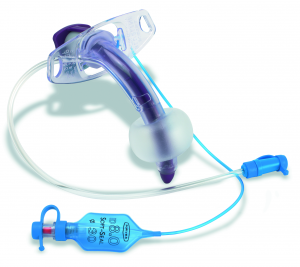
Talking tracheostomy tubes are cuffed tracheostomy tubes which have an additional tube (airflow lumen/speech lumen) that runs along the outer shaft of the tube, terminating just above the cuff, providing a lumen for air to flow. The airflow port is connected to oxygen tubing, which is connected to an external air or oxygen source.  Two to 5 liters per minute of flow is generally needed to produce a whisper type voice (Leder & Tranquina, 1989).  When increased to 5 L/min to 15 L/min, significant increases are noted, but voicing is frequently still low in volume.  Higher flows may dry the mucosa and cause discomfort.  

When the port is manually occluded, air goes through the airflow line, out the lumen above the cuff, and is directed through the vocal cords to allow speech with the cuff fully inflated.  If the patient is unable to manually occlude the port, staff or trained family members can occlude the port to enable speech. 

Advantages of talking tracheostomy tubes are that ventilator settings do not need to be adjusted because the air for speech is separate from the ventilator.  Disadvantages are that the vocal quality is frequently a soft whisper, the patient or caregiver must occlude the line for speech, the air supply line can become occluded with secretions,  discomfort and drying of the trachea from high airflow.  The patient also does not get the benefits from cuff deflation or restoring a closed system such as with a Passy-Muir Valve. 

 Home health may not provide medical air needed to use a talking tracheostomy tube and oxygen can be used instead (Pandian, V et al, 2014).

Portex® Trach-Talk™ Blue Line® Tracheostomy Tubes

The Portex Trach-Talk Blue line Tracheostomy Tubes have the additional lumen above the cuff that can be used for speech.  There is no ability to suction above the cuff or to clean the port and therefore, the lumen often clogs.  Another disadvantage is that the inner cannulas are corrugated, potentially increasing the risk of mucus plugging and difficulty in clearing secretions. (Pandian, V et al, 2014).  

Bivona® Mid-Range Aire-Cuf® and Fome-Cuf® Tracheostomy Tubes with Talk Attachment

Bivona Mid-Range Aire-Cuf and Fome-Cuf Tracheostomy Tubes with Talk Attachment also have the additional lumen above the cuff for speech.  There is no detachable thumb port or ability to suction above the cuff.  The lumen often clogs since there is no detachable port to clean the lumen.  The air cuff (Aire-Cuf) tube may be deflated with use of a speaking valve.  

Note:  The Bivona Fome-Cuf Tracheostomy Tube cannot be fully deflated and therefore is contraindicated for speaking valve use.  

Portex® Blue Line ultra® Suctionaid (BLuSA) Tracheostomy Tubes

Portex Blue Line Ultra talking tracheostomy tube

The Portex® Blue Line ultra® Suctionaid (BLuSA) Tracheostomy Tube were initially designed for above the cuff suctioning.  They are unique in that the additional lumen can be used for either suctioning or speech.  There is a thumb port that can be disconnected and used to suction secretions sitting on top of the cuff.  Subglottic suctioning reduces the amount of aspirated secretions from going into the lungs, a strategy to possibly reduce ventilator-associated pneumonia.  

Perceived advantages over other traditional talking tracheostomy tubes are that the lumen diameter for speech is larger, the inner cannula is not corrugated and the speech lumen diameter is larger.  Since the thumb port can be disconnected, the lumen for speech can be flushed with saline to keep a patent line and reduce clogging (Padian, V et al, 2014).  

Below is a video from the National tracheostomy safety project of a patient utilizing a Portex® Blue Line ultra® Suctionaid. 

Blom® Tracheostomy Tube System

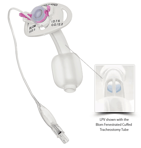
The Blom Tracheostomy Tube System is for adult individuals with a tracheostomy who weigh over 30kg (66lbs).  The system has a line of tracheostomy tubes (fenestrated/cuffed or non-fenestrated/uncuffed),  inner cannulas, Speech Cannula, Low Profile Valve (LPV), and subglottic suctioning.  The tube sizes are available in 4, 6, 8, and 10.  The fenstrated/cuffed tubes are designed for patients on mechanical ventilation while the non-fenestrated/uncuffed tubes are designed for patients breathing spontaneously.  According to the company, the fenestrations are strategically located close to the cuff in order to prevent contact with the posterior tracheal wall to reduce the risk of granulation formation in comparison to other fenestrated tracheostomy tubes.   

In brief, the Blom Tracheostomy system allows adult patients with tracheostomy the ability to produce voicing with the cuff inflated, although the cuff may also be partially or completely deflated. 

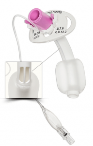
The LPV is a flap valve design (open position valve) used only for spontaneously breathing patients with the Blom Fenestrated Cuffed tube, Fenestrated Uncuffed or the Non-Fenestrated Uncuffed tube.  The cuff can be inflated, partially deflated or completely deflated.  The flap opens during inspiration and closes during exhalation, redirecting airflow through the upper airway. 

The Blom Speech Cannula is intended for patients on mechanical ventilation to speak with the cuff fully inflated.  However, the Speech Cannula can also be used with a partially deflated or completely deflated cuff.  The Speech Cannula can only to be used with the Blom Fenestrated Tracheostomy Tube.  

Blom Speech Cannula

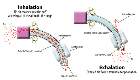
The Blom Standard Inner Cannula is removed and replaced with the Speech Cannula.  The cuff may be inflated, partially deflated or completely deflated.  During inhalation, the bubble valve expands and the flap valve opens, delivering air into the lungs.  During exhalation, the flap valve closes and the bubble valve collapses, allowing airflow through the fenestration of the tracheostomy tube and through the vocal folds and upper airway for speech.  

Speech INhale EXhale 1
Airflow with Blom Speech Cannula in place

The Blom Tracheostomy Tube System allows patients the ability to vocalize who are unable to tolerate cuff deflation.  A limitation to use of the Blom Tracheostomy Tube System is that if the resident’s tracheostomy tube is not initially placed with a Blom Tracheostomy Tube, the entire tracheostomy tube must be changed prior to using the Speech Cannula or the LPV.  Please see Pulmodyne for more information about the Blom Tracheostomy Tube System.  

Summary

The Speech-Language Pathologist plays a key role in facilitating communication and optimizing the individual’s ability to communicate effectively with clinical staff and family members. Clinicians working with these individuals should be aware of all the communication options available to provide safe and effective communication and should document how the patient best communicates their needs.  It is a critical step in restoring humanity and dignity. 

Resources:

Ten Hoorn S, Elbers PW, Girbes AR, Tuinman PR. Communicating with conscious and mechanically ventilated critically ill patients: a systematic review. Crit Care. 2016;20(1):333. Published 2016 Oct 19. doi:10.1186/s13054-016-1483-2

Khalaila R, Zbidat W, Anwar K, Bayya A, Linton DM, Sviri S. Communication difficulties and psychoemotional distress in patients receiving mechanical ventilation. Am J Crit Care 2011;20(6):470-479. 26. 

Leder SB, Traquina DN. Voice intensity of patients using a Communi-Trach I cuffed speaking tracheostomy tube. Laryngoscope 1989;99:744–747.   Medline, Google Scholar

 Pandian V, Smith CP, Cole TK, et al. Optimizing Communication in Mechanically Ventilated Patients. J Med Speech Lang Pathol. 2014;21(4):309–318.
 
Stein-Parbury J, McKinley S. Patients’ experiences of being in an intensive care unit: a select literature review. Am J Crit Care 2000; 9(1):20-27.
 
Tolotti, A, Bagnasco, A, Catania, G, Aleo, G, Pagnucci, N, Cadorin, L, Zanini, M, Rocco, G, Stievan, A, Carnevale, F, Sasso, AC. The communication experience of tracheostomy patients with nurses in the intensive care unit: A phenomenological study. Intensive and Critical Care Nursing 2018; 46(6):24-31. 


Related Posts

join trach courses

Subscribe now